 |
| Block 5: TB and occupation in South Africa - The Main Radiological Features Of Simple Silicosis |
|
| Block 5: TB and occupation in South Africa - The Main Radiological Features Of Simple Silicosis |
Simple Silicosis tends to have a Monomorphic or reproducible radiological pattern:
It is time governed, that is, it follows a time course determined by both cumulative lung silica burden and latent period of dust retention.
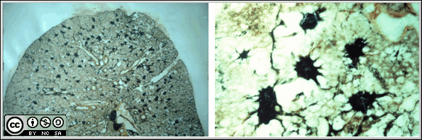
The figures above show post mortem lung section of simple, nodular silicosis, with the right hand panel being at higher magnification. The silicotic nodules appear black because of associated carbon deposition.
It is far more difficult to summarise succinctly the radiological features of Pulmonary TB. This is because PTB is a Polymorphic Disease having many, varied features, some of which may be similar to other diseases, including silicosis.
 Features such as cavitation, infiltrates, pulmonary fibrosis and distortion of intrathoracic organs occur with TB, that do not occur with silicosis, unless it is complicated by PMF or TB.
Some features seen on CXR, such as lymphadenopathy and effusions occur only with TB, and not with silicosis. Furthermore they suggest active TB disease. TB can further, on occasion, result in nodular changes in the lung, as show in the figure which shows a post-mortem section from the lung of a miner. The white nodules contain M. tuberculosis and granulonatous inflamation. Some of the commoner features of tuberculous nodulesare shown in this pathology slide. These nodules have probably occurred as a result of bronchial dissemination, rather than haematogenous dissemination (as seen in miliary TB) In the lung it can be seen that the nodules have the characteristics of:
You can see an example of peri-bronchiolar tuberculosis.
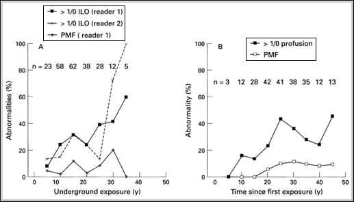
The figure (above left) presents data from a study of SA goldminers that illustrates the natural history of simple silicosis and PMF.
Simple silicosis and PMF are uncommon with less than 10 years of underground dust exposure, suggesting that this is approximately the time required to accumulate a sufficient lung dust burden to manifest silicosis. Silicosis risk and risk of PMF increases with further exposure.
The figure (above right) introduces the concept of the duration that the dust is resident in the lung, latency. Simple silicosis does not usually appear before 10 years since starting exposure whilst PMF appears 20 years after starting exposure. With longer latency, risk of silicosis and PMF increases.
The following series of slides, from Prof Ray Glyn Thomas uses x-rays to illustrate the natural history of silicosis.
Silico-tuberculosis usually refers to a worker with silicosis who develops pulmonary TB as a complication. The term also applies to workers with silicosis who additionally have stigmata of post-tb lung disease. The term does not imply any particular severity of silicosis or extent of TB.
In many instances it is readily apparent that both diseases have contributed to the radiological abnormality present. However in other instances it may be far harder to be sure about which of the two conditions is responsible for a particular abnormality and furthermore, professional opinions on the origins of some abnormalities can differ, meaning that the specificity of the CXR under such circumstances (for example, when compared to post mortem findings) can be quite low.
In the following slides the appearances of silico-tuberculosis are illustrated, with the purpose of illustration two contexts in silico-tuberculosis: 1) where both diseases are readily apparent, and 2) where both diseases are present, but both may not be readily apparent.

The slide on the left shows a post mortem lung section from a miner who had been treated for PTB. The upper pole of the lung shows irregular bands of fibrous tissue. The remainder of the lung shows dark silicotic nodules.
The following are features that may suggest TB disease activity on CXR:
If the CXR reader is suspicious about TB then the following evaluation is required:
Empirical TB treatment remains an option under certain circumstances, since "Smear negative TB" is a recognised entity. The main source of error is likely to come in immunosuppressed patients where conditions such as Cryptococcal lung disease, Kaposi’s sarcoma, viral pneumonias and otherwise unusual lung conditions are encountered.
![]()
![]()

Postgraduate Diploma in Occupational Health (DOH) - Modules 3 – 5: Occupational Medicine & Toxicology by Prof Rodney Ehrlich & Prof Mohamed Jeebhay is licensed under a Creative Commons Attribution-NonCommercial-ShareAlike 3.0 Unported License.
Permissions beyond the scope of this license may be available at http://www.healthedu.uct.ac.za/