![]()
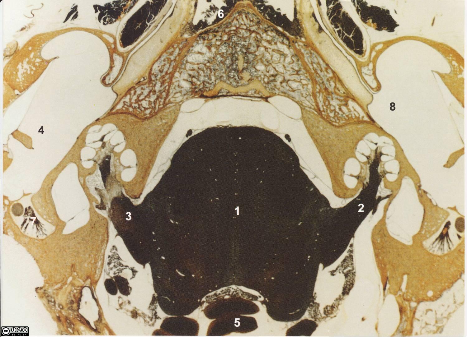
Brain stem and acoustic nerves of a cat (the left one is damaged). The cochleae are clearly visible.
The labels on the slide refer to the following:
The major dark center is the brain stem with the two eighth nerves reaching up like two arms toward the cochleas (sectioned). The right nerve is normal with the fibres stained dark. The left nerve is partly damaged by an operation and some of the nerve has been replaced by fibrous tissue.
The acoustic nerve is large, each nerve containing up to 50 000 fibres. The brown and yellow matter around the nerve is bone tissue. On both sides of the cochlea the middle ear is visible with portions of the tympanic membrane, middle ear cavity, Eustachian tube and External Auditory Meatus.
![]()